Electron Microscopy Of The Blood Of A 43 Year Old With 3 Pfizer Injections Shows Nano Razors

From Dr Hortencia Bremer
Ana Maria Mihalcea, MD, PhD | substack.com/@anamihalceamdphd
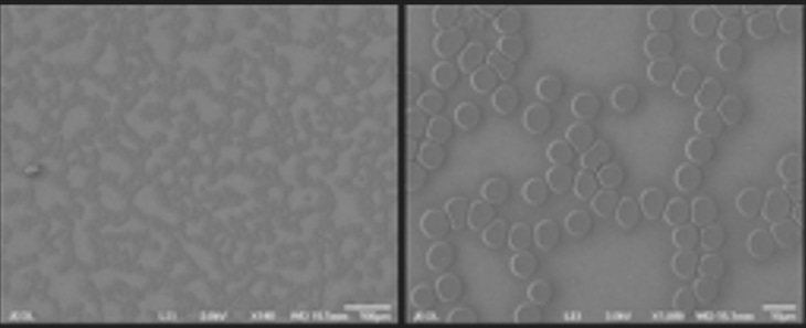
In my recent interview with Dr. Hortencia Bremer from Mexico she shared some fascinating electron microscopy images of vaccinated individuals. She kindly sent me some of these images. Dr Staninger noted that these images are consistent with these technologies, all shapes were seen by Dr. Bremer. Dr Staninger said:
“Hexagons are used for plasma pulse technology.
Triangles are part of chip series for different energy transmissions.
The nano that looked like razors are nano razors.”
This article discusses such technology:
Researchers Synthesize Nanosize Semiconductors in a Plasmonic Nanoantenna
Researchers at Hokkaido University have developed an approach to placing nanoscale semiconductors on metallic particles that is precise and cost-effective. Heat is localized on a gold nanoparticle within a butterfly-shaped nanostructure. The heat causes hydrothermal synthesis, which in turn causes semiconducting zinc oxide to crystallize on the gold nanoparticle. The Hokkaido team’s approach could open a new route to making nanosize semiconductors for nanolasing, nanolithography, and other applications.
First, the team conducted simulations to determine the optimal conditions for controlling the generation of heat in nanostructures. They used surface plasmon resonance, a process that partially converts light to heat in metallic materials. The excitation of localized surface plasmon resonances in metal nanostructures enables subwavelength photon localization and large electric field enhancement, which can then be used to enhance light-matter interactions at the nanoscale.
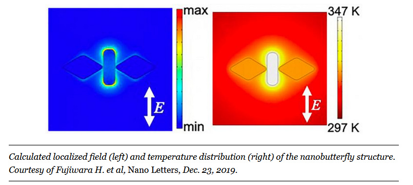
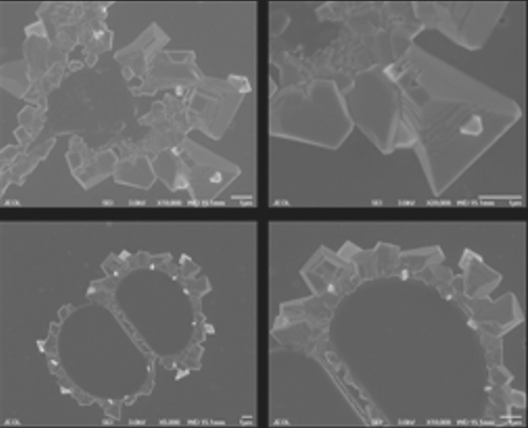
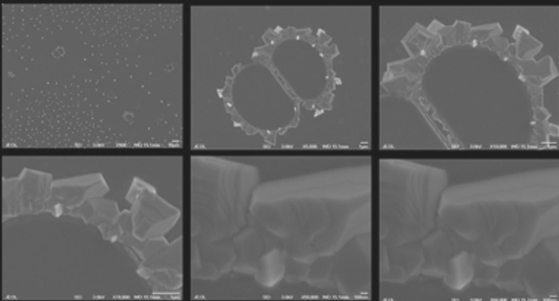
This is what the electron microscope images look like:
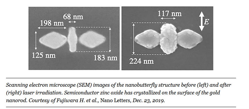
The butterfly-shaped gold nanoantennas were able to provide precise control over where plasmon-assisted hydrothermal synthesis occurred in the system and could thus enable the localized formation of nanosize semiconductors.
“Further research is expected to lead to the development of powerful nanosize light sources, highly efficient photoelectric conversion devices, and photocatalysts,” professor Keiji Sasaki said. “It could also lead to applications in semiconductor electronics and optical quantum information processing.”
The research was published in Nano Letters (www.doi.org/10.1021/acs.nanolett.9b04073).
Here are further images of the blood, and you can see these tiny razors in the background:
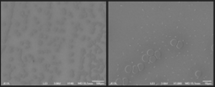
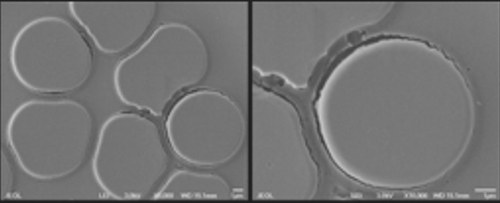
Nanorazors visible compared to red blood cell that is approx. 5 micrometer in diameter. This very much reminds me of Chemist Andreas Noack who talked about the Graphene being like razorblades to the vascular endothelium. [See | Related: Article link at bottom of article]
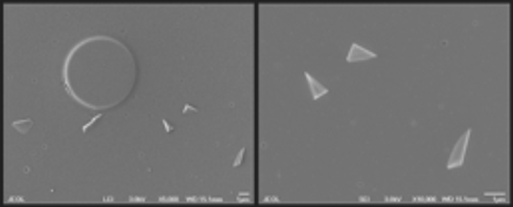
Here are higher magnification:
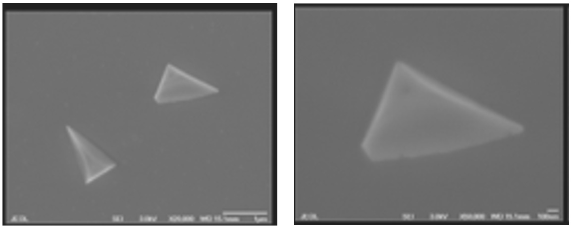
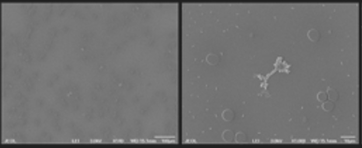

Note that the round structures look like the liposomal bubbles we have been seeing, but we had no diea what is on the surface.
It makes me wonder if some of the microbubbles we see are laden with these nanorazor particles. You can see
Microbubbles Are Microrobots That Build Microchips - Correlation with COVID19 Microscopy Findings
Summary:
I have a lot more images that I will post as well. Such nanorazors could significantly contribute to injury and micro clotting. If these structures are plasmonic antennas the frequency emission can cause cellular damage. They look very similar to the inorganic nanoparticles that Dr Antoinetta Gatti found in childhood vaccines and she showed their presence in leukemia patients. I have been seeing a large volume of unvaccinated patients with turbo cancers that has exponentially increased - in young patients. Many when they receive chemo have rapid recurrences to stage 4 disseminated disease. Given multiple recent articles I have posted showing the association of nanoparticles and cancer, I encourage colleagues to look at those associations.
This is the article by Dr. Antoinetta Gatti:
Background: Continuing growth in the incidence of leukemia suggests a possible environmental etiology correlated to the increase of environmental pollution. Recently, environmental particulate pollution (EPP) has been declared by IARC a Class I carcinogenic agent; it looks reasonable to presume that not only chemicals like benzene and its derivatives, but also other components like EPP could be worth of study. No specific researches have up to now focused the role of EPP on acute myeloid leukemia; we thus have identified a suitable instrumentation and protocol to show the presence and composition of particulate matter in blood samples of patients affected by acute myeloid leukemia patients and in healthy controls.
Methods: 38 peripheral blood samples (19 acute myeloid leukemia, 19 healthy controls) were analyzed by means of an Environmental Scanning Electron Microscopy (ESEM) coupled with an Energy Dispersive Spectroscopy (EDS) a sensor capable of identifying the composition of micro- and nano-particles of exogenous nature in pathological tissues (applied for the first time in the current study on blood samples). The results were statistically treated with unpaired two-tailed Student's t-test, MANOVA and Principal Component Analysis.
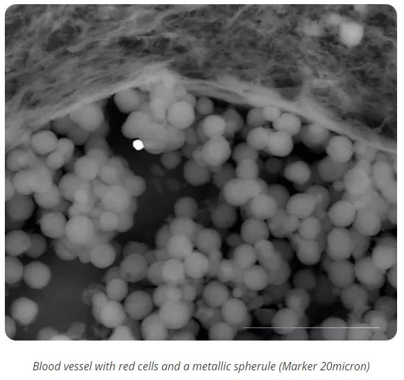
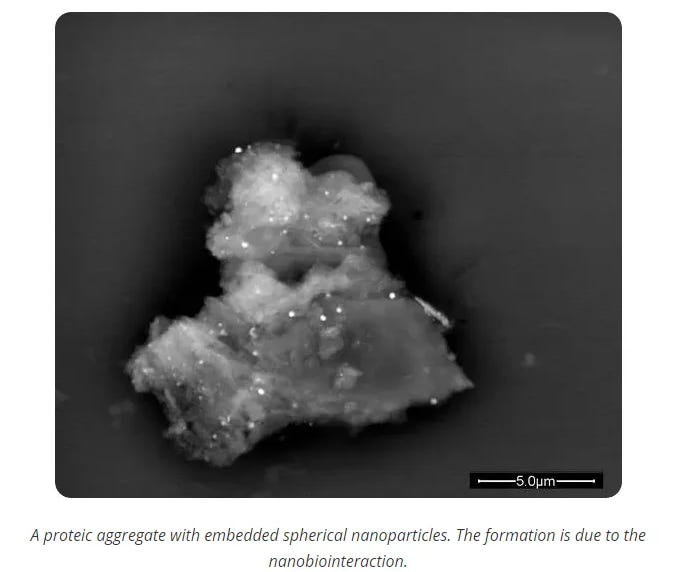
Results: A consistent quantity of micron-, submicron- and nano-sized foreign bodies (from 20 micron down to 100nm) was documented in 18/19 AML cases, whereas they were absent or rare in the controls. The particles appeared as singlet and aggregates (ranging from 5 to 20micron), either in close contact with blood elements or interacting with plasma. Some reacted with blood proteins thus forming composite clusters. A total of 141 aggregates (median 8, range 0-18) in AML, compared to a total of 12 aggregates in controls (median 1, range 0-3) were counted. The aggregate analysis showed variable sizes and number of particles, with a total of 5394 particles in leukemia cases compared to a total of 207 in controls. The total numbers of aggregates and particles were statistically different between cases and controls (MANOVA, P<0.001 e P=0.009 respectively). Aggregates were then analyzed with EDS, identifying their elemental composition. The particles mostly contained highly reactive metals, and appeared not biocompatible and not biodegradable. In particular, micro- and nano-sized particulates were segregated in organic-inorganic clusters, with statistically higher frequency of a subgroup of elements in AML samples (Si, P=0.03; Al, P=0.03; Fe, P=0.002; Ti, P=0.04, Cu, P=0.02, respectively). The analyses of the chemical spectra in some cases allowed to recognize and identify the source of the contamination.
Conclusion: In conclusion, we demonstrated the exposure of a subset of AML patients to environmental contaminants, with invasive character in the human body, not biocompatible and biopersistent. AML, as well as myelodysplastic syndromes, are derived from precursor cells critical in innate immunity, that submicronic particles could have triggered. New etiopathogenic hypotheses involving an interaction among sub-micron and nanosized particles with blood components are under evaluation.
Of course she was the one to also confirm these metallic toxic nanoparticles in 44 childhood vaccines:
New quality-control investigations on vaccines: micro- and nanocontamination
Here are some of her images: Nanopathology
Transhuman Vol 2 and Light Medicine contain combined over 100 pages of how to remove heavy metal nanoparticles with the use of EDTA Chelation and case studies.
Light Medicine explains how these metals scramble the biophotonic light emitted by our DNA and other cellular structures that produce the holographic genetic encoding of who we are according to Dr. Peter Garaiev’s gene wave model and Dr. Fritz Alexander Popp. All carcinogens interfere with the mechanism of biophotonic cellular self repair by creating incoherence in the emission spectrum.
Original Article: https://anamihalceamdphd.substack.com/p/electron-microscopy-of-the-blood
Note: Comments placed in [ ] are added by Truth11.com editor. For example; [Flu]
Related:


Truthful comments that add value to our readers, such as additional information, clarification, validation or worthy rebuttal will be posted. The goal of comments on articles is to gain additional truth.

Support Truth11.com With A $1, $3 or $11 Monthly Subscription
Thank you for supporting independent media. Your subscription is directly helping us cover costs to keep Truth11.com going! Thank you Truth Warriors!


Comments ()